Dermatopathology
Wiley-Blackwell (Verlag)
978-1-119-14945-3 (ISBN)
- Titel ist leider vergriffen;
keine Neuauflage - Artikel merken
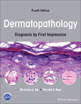
The atlas that helps you differentiate visually similar diseases Written with the dermatology trainee in mind, Dermatopathology: Diagnosis by First Impression uses more than 800 high resolution color images to introduce a simple and effective way to defuse the confusion caused by dermatopathology slides. Focused on commonly tested entities, and using low- to high-power views, this atlas emphasizes the key differences between visually similar diseases by using appearance as the starting point for diagnosis. The Third Edition provides: *800 high resolution and annotated photographs, now all fully downloadable *'Key Differences' to train the eye on distinctive diagnostic features * Disease-based as well as alphabetical indexes *75 new interactive self-assessment questions to perfect your diagnostic skills * Brand new algorithms for pattern analysis Dermatopathology: Diagnosis by First Impression, Third Edition, once again provides simple and effective guidance to help you approach dermatopathology and accurate diagnosis of skin disease.
Christine J. Ko is a Professor of Dermatology and Pathology at Yale University School of Medicine. She trained in dermatology at University of California, Irvine, where she was strongly influenced by Dr. Barr. She subsequently completed a fellowship in dermatopathology under Dr. Scott Binder at University of California, Los Angeles. She lectures nationally and internationally; and has published numerous book chapters, journal articles, and five textbooks/atlases in the fields of dermatology and dermatopathology. Ronald J. Barr is Professor Emeritus of Dermatology and Pathology at the University of California, Irvine. He is a nationally and internationally recognized dermatopathologist with board certification in dermatology, anatomic pathology, and dermatopathology. He received the Founders' Award from the American Society of Dermatopathology for his myriad contributions to the field of dermatopathology and the Society's Walter Nickel Award for excellence in teaching dermatopathology. He has authored over 150 original articles and book chapters. He is also past president of the American Society of Dermatopathology and past president of the American Board of Dermatology.
Preface, vi Acknowledgments, vii About the Companion Website, viii Introduction, 1 Chapter 1 Shape on Low Power, 23 Epidermis Regular acanthosis, 25 Lobular proliferation, 29 Reticulated proliferation, 35 Central pore, 42 Epidermal perforation, 46 Dermis Circular islands, 49 Cords/tubules and comma shapes, 53 Space with a lining, 59 Papillations, 70 Polypoid (dome-shaped), 77 Square/rectangular, 82 Palisading reactions, 88 Pseudoepitheliomatous hyperplasia above abscesses, 93 Pink ball, (see Chapter 6) Chapter 2 Gestalt: Rash/inflammatory, 97 Epidermal changes Parakeratosis, 99 Spongiosis, 102 Papulosquamous (psoriasiform), 106 Interface (vacuolar), 112 Interface (lichenoid), 117 Inflammation: Specific patterns and Cell Type Epidermal eosinophils, 123 Perivascular, 127 Band-like dermal/papillary dermal infiltrate, 131 Diffuse/nodular, 137 Subcutaneous, 144 Chapter 3 Cell Type, 153 Melanocytic, 155 Spindle cells, 164 Endothelial, 178 Giant, 192 Clear, 202 Chapter 4 Top-Down , 219 Hyperkeratosis/parakeratosis, 221 Upper epidermal change, 228 Acantholysis, 238 Subepidermal space/cleft, 248 Granular material in cells, 255 Busy dermis, 260 Dermal material, 263 Fat necrosis, 276 Chapter 5 Color Blue, 279 Blue tumor, 281 Mucin and glands or ducts, 291 Mucin, 295 Chapter 6 Color Pink, 303 Pink ball of spindle cells, 305 Pink material, 308 Pink dermis, 315 Epidermal necrosis, 317 Index (Pattern), 323 Index (Histological Category), 329 Index (Alphabetical), 333
| Erscheinungsdatum | 25.10.2016 |
|---|---|
| Verlagsort | Hoboken |
| Sprache | englisch |
| Maße | 217 x 283 mm |
| Gewicht | 1016 g |
| Themenwelt | Medizin / Pharmazie ► Medizinische Fachgebiete ► Dermatologie |
| Studium ► 2. Studienabschnitt (Klinik) ► Pathologie | |
| ISBN-10 | 1-119-14945-2 / 1119149452 |
| ISBN-13 | 978-1-119-14945-3 / 9781119149453 |
| Zustand | Neuware |
| Informationen gemäß Produktsicherheitsverordnung (GPSR) | |
| Haben Sie eine Frage zum Produkt? |
aus dem Bereich