Clinical Radiology of the Horse (eBook)
John Wiley & Sons (Verlag)
978-1-118-91226-3 (ISBN)
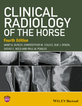
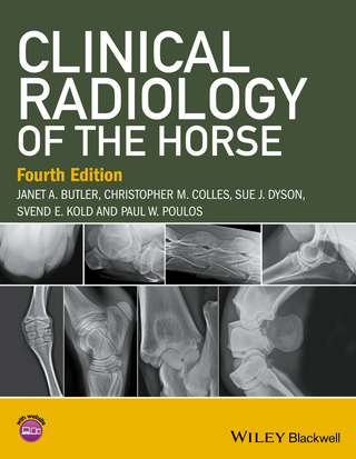
Clinical Radiology of the Horse is the best-selling, practical guide to all areas of equine radiography and radiology written by an experienced group of clinicians with a broad range of backgrounds.
- Offers an atlas of normal and clinical images, as well as a comprehensive guide to techniques, equipment, positioning, and interpretation for general veterinary practitioners and specialists in imaging and orthopaedics
- Updates to this fourth edition fully reflect the move to digital imaging with many new figures in the book and major revisions to the chapters on the head, thorax, and abdomen
- Contains expanded coverage of the foot, pastern, and fetlock (now in separate chapters)
- Includes a password-protected website with all the images from the book as well as over 200 additional images with examples of more subtle lesions, more fractures, correct technique and positioning versus incorrect, immature horses, progression of disease, and pathological images
Janet A. Butler
Jan specialises in equine radiography and has 40 years' experience in this field. She joined the Animal Health Trust in Newmarket, UK in 1975 where she gained considerable experience working with many internationally renowned veterinary surgeons. Since 1997 she has been working in private practice, initially at the Willesley Equine Clinic, UK, which since 2009 has been part of the B&W Equine Group.
Christopher M. Colles
Chris qualified from the Royal Veterinary College, UK in 1971. After three years in mixed practice (where he obtained a Part I Diploma in Radiology) he joined the Animal Health Trust as a clinician in 1975. He has carried out research in many areas of equine orthopaedics and radiology, having a particular interest in the horse's foot. In 1988 he returned to practice, where he became a senior partner in Avonvale Veterinary Practice, specialising in equine orthopaedics, until his recent retirement from practice. He is recognised by the Royal College of Veterinary Surgeons as a Specialist in Equine Orthopaedic Surgery. Chris was awarded an Honorary Fellowship of the Worshipful Company of Farriers in 2000 in recognition of his research into conditions of the foot, and involvement with farriery education.
Sue J. Dyson
After qualifying from the University of Cambridge in 1980, Sue worked for a year at New Bolton Center, University of Pennsylvania, and then spent a year in private practice in Pennsylvania. Sue then joined the Centre for Equine Studies of the Animal Health Trust, UK, where she has specialised in lameness diagnosis and diagnostic imaging. Sue is recognised as a Specialist in Equine Orthopaedics by the Royal College of Veterinary Surgeons and holds the RCVS Diploma in Equine Orthopaedics. She is an Associate of the European College of Veterinary Diagnostic Imaging. She has published widely on lameness, radiography ultrasonography, nuclear scintigraphy and magnetic resonance imaging.
Svend E. Kold
Svend qualified from The Royal Veterinary and Agricultural University in Copenhagen in 1979. He then spent over 10 years at the Animal Health Trust in Newmarket. After a sabbatical year at Colorado State University, he joined the Willesley Equine Clinic, UK, where he was a partner until 2009. He is now a private consultant. He specialises in lameness and orthopaedic diagnostics and surgery and is recognized as a Specialist in Equine Orthopaedic Surgery by the Royal College of Veterinary Surgeons. He is veterinary consultant to a European equine insurance company and is involved as an expert in many equine legal cases, both in the UK and Europe. He has published regularly on orthopaedic subjects.
Paul W. Poulos
Following graduation from the University of California at Davis in 1960, Paul founded a private practice. In 1972 he returned to Davis to specialise in radiology where he was became Diplomate of the American College of Veterinary Radiology. He moved to the Royal Veterinary College of Stockholm, Sweden and later was Associate Professor at Radiology at the University of Utrecht. On return to the USA, was Professor of Radiology at University of Florida, and later chairman of the Department of Radiology. In 1990 Paul left academia to establish his own consulting practice, Poulos Veterinary Imaging, based in Ukiah, California. He has published widely on osteochondrosis, navicular disease and diseases of the fetlock. Paul is now retired and has not contributed to the fourth edition of the book.
With contributions from Sarah Puchalski
Following graduation from the University of Saskatchewan in 1999, Sarah worked for two years at New Bolton Center, University of Pennsylvania. In 2001 she moved to the University of Davis, California to specialise in diagnostic imaging. In 2004, she was awarded Diplomate of the American College of Veterinary Radiology. After 12 years in the diagnostic imaging department of the University of Davis, California, Sarah recently moved into private practice in California, but remains Adjunct Associate Professor of Diagnostic Imaging, Department of Surgical and Radiological Sciences, School of Veterinary Medicine, University of California, Davis. Sarah has published widely on many aspects of diagnostic imaging.
Clinical Radiology of the Horse is the best-selling, practical guide to all areas of equine radiography and radiology written by an experienced group of clinicians with a broad range of backgrounds. Offers an atlas of normal and clinical images, as well as a comprehensive guide to techniques, equipment, positioning, and interpretation for general veterinary practitioners and specialists in imaging and orthopaedics Updates to this fourth edition fully reflect the move to digital imaging with many new figures in the book and major revisions to the chapters on the head, thorax, and abdomen Contains expanded coverage of the foot, pastern, and fetlock (now in separate chapters) Includes a password-protected website with all the images from the book as well as over 200 additional images with examples of more subtle lesions, more fractures, correct technique and positioning versus incorrect, immature horses, progression of disease, and pathological images
Title Page 3
Copyright Page 4
Contents 5
About the authors 7
Preface to the fourth edition 9
About the companion website 11
Chapter 1 General principles 13
INTRODUCTION 13
PRINCIPLES OF RADIOGRAPHY 15
Production of x-rays 15
Production of a radiographic image 16
Exposure factors 16
X-ray film and image intensifying screens 17
Film processing 18
Radiographic practice 20
Radiation safety 24
Examination for purchase 25
Records and labelling 26
PRINCIPLES OF RADIOGRAPHIC INTERPRETATION: RADIOLOGY 27
RADIOLOGICAL APPEARANCE OF PHYSIOLOGICAL CHANGES AND SOME COMMON PATHOLOGICAL LESIONS 32
Bone changes 32
Bone lesions 36
Joint lesions 44
Additional figures 50
FURTHER READING 51
Chapter 2 Computed and digital radiography 53
COMPUTERISED RADIOGRAPHY 53
Imaging plate reading 54
DIRECT DIGITAL RADIOGRAPHY 55
IMAGE RESOLUTION 55
EXPOSURE FACTORS 56
Overexposure 56
Underexposure 57
Algorithms 57
MONITORS 58
COMPUTED RADIOGRAPHY ARTEFACTS 59
Imaging plate artefacts 59
Plate reader artefacts 59
Operator-induced artefacts 61
IMAGE READING 62
Image manipulation 62
IMAGE ARCHIVING AND TRANSMISSION 64
ADVANTAGES OF DIGITAL RADIOGRAPHY COMPARED WITH CONVENTIONAL FILM– SCREEN RADIOGRAPHY 65
Additional figures 66
FURTHER READING 66
Chapter 3 The foot 67
Distal phalanx (pedal bone) 67
RADIOGRAPHIC TECHNIQUE 67
Equipment 67
Positioning 68
NORMAL ANATOMY 74
Immature horse 74
Skeletally mature horse 74
NORMAL VARIATIONS AND INCIDENTAL FINDINGS 82
SIGNIFICANT FINDINGS 88
Pedal osteitis 88
Osseous cyst-like lesions 92
Keratoma 93
Tumours 95
Ossification of the ungular cartilages (sidebone) 95
Entheseophytes adjacent to the extensor process of the distal phalanx 100
Osseous changes at the insertion of the deep digital flexor tendon and distal sesamoidean impar ligament 100
Degenerative joint disease of the distal interphalangeal joint 100
Subluxation of the distal interphalangeal joint 103
Agenesis or hypoplasia of the distal phalanx 104
Fractures 104
Hoof 109
RADIOGRAPHIC TECHNIQUE 109
Technique to assess hoof balance 110
NORMAL ANATOMY 110
NORMAL VARIATIONS AND INCIDENTAL FINDINGS 111
DIGITAL ANGIOGRAPHY AND VENOGRAPHY 112
SIGNIFICANT FINDINGS 112
Hoof balance 112
SIGNIFICANT FINDINGS 114
Laminitis 114
Long-toe low-heel syndrome 120
Mediolateral foot imbalance 123
Infection 123
Penetrating injuries 124
Hoof wall separation 124
Navicular bone 125
RADIOGRAPHIC TECHNIQUE 125
Equipment 125
Positioning 126
NORMAL ANATOMY 131
Immature horse 131
Skeletally mature horse 131
NORMAL VARIATIONS AND INCIDENTAL FINDINGS 138
SIGNIFICANT FINDINGS 139
Common artefacts 139
Congenital abnormalities of the navicular bone 140
Navicular disease 140
New bone formation 147
Mineralisation in the deep digital flexor tendon 151
Infection 151
Fractures 152
Proximal displacement of the navicular bone 153
Osseous lesions of the foot which may be missed using radiography 154
Additional figures 154
FURTHER READING 155
Chapter 4 The proximal and middle phalanges and the proximal interphalangeal joint 161
RADIOGRAPHIC TECHNIQUE 161
Equipment 161
Positioning 161
NORMAL ANATOMY 162
Immature horse 162
Skeletally mature horse 162
NORMAL VARIATIONS AND INCIDENTAL FINDINGS 165
SIGNIFICANT FINDINGS 169
Dysplasia of the proximal interphalangeal joint 170
Subluxation of the proximal interphalangeal joint 172
Osseous cyst-like lesions 172
Degenerative joint disease of the proximal interphalangeal joint 173
New bone formation 174
‘Scalping’ injury of the proximal or middle phalanx 178
Collateral ligament injury of the proximal interphalangeal joint 178
Rupture of the straight sesamoidean ligament 178
Subchondral bone trauma of the proximal axial aspect of the proximal phalanx 178
Subchondral bone trauma of the proximal interphalangeal joint 178
Subchondral bone trauma of the distal dorsal aspect of the middle phalanx 179
Fractures 179
Dystrophic mineralisation 183
Tumours 183
Additional figures 184
FURTHER READING 184
Chapter 5 Metacarpophalangeal and metatarsophalangeal (fetlock) joints 187
RADIOGRAPHIC TECHNIQUE 187
Equipment 187
Positioning 187
NORMAL ANATOMY 193
Immature horse 193
Skeletally mature horse 194
NORMAL VARIATIONS AND INCIDENTAL FINDINGS 199
SIGNIFICANT FINDINGS 201
Soft-tissue swelling 201
Degenerative joint disease 205
Subchondral bone trauma 208
Osteochondrosis, developmental orthopaedic disease and osteochondral fragments 208
Physitis 211
Osseous cyst-like lesions 211
Sesamoiditis 211
Infectious osteitis of the palmar or abaxial aspect of a proximal sesamoid bone 215
Infectious and traumatic osteitis of the axial aspect of the proximal sesamoid bones 215
Osseous cyst-like lesion in the proximal aspect of a proximal sesamoid bone 216
Abnormal position of the proximal sesamoid bones 216
Luxation 216
Stress-related bone injury 216
Fractures 217
Additional figures 221
Further reading 221
Chapter 6 The metacarpal and metatarsal regions 227
RADIOGRAPHIC TECHNIQUE 227
Lateromedial, dorsopalmar and oblique views 227
Dorsoproximal-palmarodistal oblique views 229
Other imaging techniques 230
NORMAL RADIOGRAPHIC ANATOMY: ITS VARIATIONS AND INCIDENTAL FINDINGS 231
Lateromedial view 231
Dorsopalmar view 233
Dorsolateral-palmaromedial oblique and dorsomedial?palmarolateral oblique views 233
SIGNIFICANT RADIOLOGICAL ABNORMALITIES 241
Periostitis 241
Infectious osteitis and osteomyelitis 251
Angular limb deformities originating from the diaphysis of the third metacarpal or metatarsal bone 251
Physitis of the third metacarpal bone 253
Mineralisation in the soft tissues 254
Hypertrophic osteopathy 254
Enostosis-like lesions and panosteitis 254
Fractures 254
Tumours 264
Additional figures 264
FURTHER READING 267
Chapter 7 The carpus and antebrachium 271
RADIOGRAPHIC TECHNIQUE 271
Equipment 271
Positioning for the carpus 271
Positioning for the antebrachium (radius) 273
NORMAL ANATOMY 274
Immature horse 274
Skeletally mature horse 274
NORMAL VARIATIONS AND INCIDENTAL FINDINGS 283
SIGNIFICANT FINDINGS 287
Soft-tissue swelling 287
Intercarpal ligament desmitis 290
Degenerative joint disease 290
New bone formation 294
Sclerosis of the third carpal bone 297
Osseous cyst-like lesions 299
Polydactyly 300
Physitis 300
Carpal angular limb deformities 301
Incomplete carpal ossification 303
Osteochondroma 303
Carpal subluxation 305
Carpal fractures 305
Additional figures 310
FURTHER READING 310
Chapter 8 The shoulder, humerus, elbow and radius 313
Scapulohumeral (shoulder) joint and humerus 313
RADIOGRAPHIC TECHNIQUE 313
Equipment 313
Positioning 313
RADIOGRAPHIC ANATOMY, NORMAL VARIATIONS AND INCIDENTAL FINDINGS 317
Birth to 3 years old 317
Skeletally mature horse 318
Arthrography 324
SIGNIFICANT RADIOLOGICAL ABNORMALITIES 325
Osteochondrosis 325
Osseous cyst-like lesions 327
Degenerative joint disease 331
Mineralisation in the tendon of biceps brachii 332
Lesions of the humeral tubercles 333
Congenital abnormalities of the bicipital apparatus 333
Heterotopic ossification 334
Abnormalities of the scapulohumeral joint in Shetland Ponies and Miniature Horses 334
Infection 335
Luxation of the scapulohumeral joint 337
Enostosis-like lesions 338
Fractures 338
Bone fragility syndrome 341
Humeroradial, humeroulnar and radioulnar (elbow or cubital) joints and radius 342
RADIOGRAPHIC TECHNIQUE 342
Equipment 342
Positioning 342
RADIOGRAPHIC ANATOMY, NORMAL VARIATIONS AND INCIDENTAL FINDINGS 343
Birth to 3 years old 343
Skeletally mature horse 343
SIGNIFICANT RADIOLOGICAL ABNORMALITIES 347
Osteochondrosis 347
Osseous cyst-like lesions 347
Degenerative joint disease 347
Periosteal proliferative reactions (enthesopathy) at the site of insertion of biceps brachii on the radial tuberosity 351
Entheseous new bone at the sites of attachment of the collateral ligaments of the humeroradial joint 351
Periosteal reaction at the site of origin of the accessory ligament of the superficial digital flexor tendon 353
Luxation of the elbow joint 353
Infection 354
Osteochondroma of the distal aspect of the radius 354
Hereditary multiple exostosis 354
Hypertrophic osteopathy 354
Fractures (Figure 8.39 see also Figure 8.27)
Tumours 358
Additional figures 358
FURTHER READING 358
Chapter 9 The tarsus 361
RADIOGRAPHIC TECHNIQUE 361
Equipment 361
Positioning 361
RADIOGRAPHIC ANATOMY: NORMAL VARIATIONS AND INCIDENTAL FINDINGS 364
Immature horse 364
Skeletally mature horse 365
SIGNIFICANT RADIOLOGICAL ABNORMALITIES 380
Congenital abnormalities 380
Tarsal bone collapse 381
Osteochondrosis 382
Physitis or physeal dysplasia 386
Degenerative joint disease 387
Mineralisation of the central or third tarsal bone 395
Bone trauma 395
Periosteal proliferative reactions at the sites of attachment of ligaments and joint capsules 395
Fragmentation of the proximal tubercle of the talus 398
Luxation 399
Distension of the tarsal sheath and synoviocoeles (Thoroughpin) 399
Infectious arthritis and osteomyelitis 401
Osseous cyst-like lesions 402
Hypertrophic osteopathy 404
Fractures 404
Tumours 407
Additional figures 407
FURTHER READING 407
Chapter 10 The stifle and tibia 411
Stifle 411
RADIOGRAPHIC TECHNIQUE 411
Equipment 411
Positioning 412
NORMAL ANATOMY, VARIATIONS AND INCIDENTAL FINDINGS 415
Immature horse 415
Skeletally mature horse 417
SIGNIFICANT FINDINGS 426
Osteochondrosis 426
Osseous cyst-like lesions 433
Necrosis of the femoral condyles 438
Bone trauma 438
Physitis 439
Degenerative joint disease 439
Infection 441
Miscellaneous soft-tissue injuries 442
Patellar luxation 447
Infectious osteitis of the patella 448
Chondromalacia of the patella 449
Fractures 449
Tibia 452
RADIOGRAPHIC TECHNIQUE 452
NORMAL ANATOMY, VARIATIONS AND INCIDENTAL FINDINGS 453
SIGNIFICANT RADIOLOGICAL ABNORMALITIES 454
Enostosis-like lesions and other focal opacities 454
Fractures 455
Additional figures 456
FURTHER READING 457
Chapter 11 The head 461
RADIOGRAPHIC TECHNIQUE 461
Equipment 461
GENERAL POSITIONING OF THE PATIENT 462
Cranium 463
RADIOGRAPHIC TECHNIQUE 464
NORMAL ANATOMY, VARIATIONS AND INCIDENTAL FINDINGS 464
Immature horse 464
Skeletally mature horse 464
SIGNIFICANT FINDINGS 468
Dentigerous cysts 468
Choanal restriction or atresia 468
Abnormalities of the nasal septum 469
Ethmoid haematoma and diseases of the Eustachian tube diverticulum (guttural pouch) 469
Temporohyoid osteoarthropathy 469
Nasal polyps 473
Hydrocephalus 473
Entheseophyte formation on the occiput associated with the nuchal ligament 473
Fractures 475
Paranasal sinuses (frontal, maxillary, conchal) and maxilla 478
RADIOGRAPHIC TECHNIQUE 478
NORMAL ANATOMY, VARIATIONS AND INCIDENTAL FINDINGS 479
SIGNIFICANT FINDINGS 479
Sinusitis 479
Submucosal cyst 484
Maxillary sinus cyst 484
Maxillary cysts 486
Ethmoid haematoma 486
Other causes of opacity of the maxillary sinus 486
Increased lucency of the maxillary sinus 491
Cyst-like lesions of the incisive bone (premaxilla) 492
Teeth and mandible 492
RADIOGRAPHIC TECHNIQUE 494
Mandible 494
Temporomandibular joint 494
Cheek teeth 496
Incisors 499
NORMAL ANATOMY AND VARIATIONS 500
SIGNIFICANT FINDINGS 501
Brachygnathia and prognathia 501
Polydontia 501
Oligodontia 501
Tumours of dental origin 505
Equine odontoclastic tooth resorption and hypercementosis (EOTRH) 505
Dental fractures 507
Tooth root infection 511
Periodontal disease 513
Disorders of the erupted crowns 513
Mandibular periostitis 515
Osteomyelitis 516
Mandibular cysts 516
Craniomandibular osteopathy 517
Tumours 517
Temporomandibular joint degenerative joint disease 518
Luxation of the temporomandibular joint 520
Fractures 520
Sequestrum of the interdental space 522
Pharynx, larynx and Eustachian tube diverticulum 524
RADIOGRAPHIC TECHNIQUE 524
NORMAL ANATOMY, VARIATIONS AND INCIDENTAL FINDINGS 525
SIGNIFICANT FINDINGS 528
Eustachian tube diverticulum empyema 528
Eustachian tube diverticulum chondrosis 530
Eustachian tube diverticulum tympany 530
Eustachian tube diverticulum mycosis 532
Eustachian tube diverticulum masses 532
Pharyngeal lymphoid hyperplasia 532
Fracture of the hyoid apparatus 532
Epiglottic entrapment 533
Epiglottic shortening 534
Sub-epiglottic cysts 535
Arytenoid chondritis 535
Other laryngeal disorders 537
Dorsal displacement of the soft palate 537
Cleft palate 538
Multilobular osteoma (chondroma rodens) 539
Aneurysmal bone cyst 539
Additional figures 539
FURTHER READING 539
Chapter 12 The vertebral column 543
Cervical vertebrae 543
RADIOGRAPHIC TECHNIQUE 543
Equipment 543
Positioning 544
NORMAL ANATOMY, VARIATIONS AND INCIDENTAL FINDINGS 546
Lateral-lateral images 546
Characteristics of the cervical vertebrae 553
Lateroventral-laterodorsal images 557
SIGNIFICANT RADIOGRAPHIC ABNORMALITIES 561
Congenital abnormalities 561
Developmental abnormalities 561
Modelling of the cervical articular process joints 567
Subluxation of the atlantoaxial joint 572
Subluxation of the caudal cervical and first thoracic vertebrae 573
Entheseophyte formation on the occiput associated with the nuchal ligament 573
Degenerative changes of the intercentral articulations 574
Osteomyelitis 575
Discospondylitis 575
Osseous cyst-like lesions 576
Neoplasia 577
Soft-tissue lesions 578
Spondylosis 578
Scoliosis 579
Fractures 579
Thoracolumbar vertebrae 581
RADIOGRAPHIC TECHNIQUE 581
Equipment 581
Positioning 582
NORMAL ANATOMY, VARIATIONS AND INCIDENTAL FINDINGS 583
Immature horse 583
Skeletally mature horse 583
SIGNIFICANT FINDINGS 598
Sprain of the supraspinous ligament 598
Interspinous ligament enthesopathy 599
Impingement and overriding of the spinous processes 599
Other abnormalities of the spinous processes 605
Vertebral fusion 605
Lordosis, kyphosis and scoliosis 606
Degenerative joint disease 606
Ossifying spondylosis (spondylosis deformans) 609
Infectious osteitis 611
Discospondylitis 611
Luxation 612
Fractures 612
Neoplasia 612
Sacrum and coccygeal vertebrae 614
Additional figures 616
FURTHER READING 616
Chapter 13 The pelvis and femur 621
Pelvis 621
RADIOGRAPHIC TECHNIQUE 621
Equipment 621
Positioning 623
NORMAL ANATOMY, VARIATIONS AND INCIDENTAL FINDINGS 625
Immature horse 625
Skeletally mature horse: ventrodorsal views 626
Skeletally mature horse: lateral 300 dorsal-lateroventral oblique views 634
SIGNIFICANT FINDINGS 634
Hip dysplasia 634
Subluxation of the coxofemoral joint 634
Luxation of the coxofemoral joint 636
Infectious arthritis/osteomyelitis 636
Degenerative joint disease 636
Sacroiliac joint disease 638
Osseous cyst-like lesions 638
Osteochondrosis 638
Osteochondroma of the pubis 640
Fractures 640
Femur 644
RADIOGRAPHIC TECHNIQUE 644
Equipment 644
Positioning 645
RADIOGRAPHIC ANATOMY 645
RADIOGRAPHIC ABNORMALITIES 646
Enostosis?like lesions 646
Fractures of the femur 646
FURTHER READING 648
Chapter 14 The thorax 651
RADIOGRAPHIC TECHNIQUE 651
Equipment 651
Positioning 651
Other imaging techniques 653
Monitoring pulmonary and pleural disease 656
NORMAL ANATOMY 657
Immature horse 662
Mature horse 662
NORMAL VARIATIONS AND INCIDENTAL FINDINGS 663
Factors influencing interpretation 663
SIGNIFICANT FINDINGS 664
Patterns of lung disease 664
Diseases of the pleural space and mediastinum 0
Diseases of the lung 0
Additional figures 0
FURTHER READING 0
Chapter 15 The alimentary and urinary systems 699
RADIOGRAPHIC TECHNIQUE 699
Equipment 699
Positioning 700
Contrast examinations 700
Oesophagus 706
RADIOGRAPHIC TECHNIQUE 706
Contrast examination of the oesophagus 706
DISEASES OF THE OESOPHAGUS 707
Diseases that decrease the diameter 707
Diseases that increase the diameter 710
Oesophageal dysfunction and obstruction (choke) 713
Abdomen and gastrointestinal tract 717
RADIOGRAPHIC ANATOMY 717
Foal abdomen 717
Adult abdomen 718
ANATOMICAL VARIATIONS 720
Contrast studies in the foal 720
DISEASES OF THE GASTROINTESTINAL TRACT 720
Small intestinal obstruction 720
Large intestinal obstruction 721
Atresia coli 723
Ileocolonic aganglionosis 723
Gastroenteritis 723
Gastroduodenal ulcer disease in foals 723
Rupture of a hollow viscus 726
Enterolithiasis of adult horses 727
Sand impaction 727
Urinary system 727
CONTRAST EXAMINATION 727
DISEASES OF THE URINARY BLADDER OF THE FOAL 729
Patent urachus 729
Cystitis 729
Rupture 729
Additional figures 732
FURTHER READING 732
Chapter 16 Miscellaneous techniques 735
ARTHROGRAPHY AND BURSOGRAPHY 735
Technique 736
Diagnostic criteria 736
TENDONOGRAPHY 736
Technique and diagnostic criteria 737
ANGIOGRAPHY 737
Technique 738
Diagnostic criteria 739
VENOGRAPHY 742
Technique and interpretation 743
MYELOGRAPHY 745
Technique 745
Interpretation of the myelogram 746
Diagnostic criteria 747
PNEUMOCYSTOGRAPHY 755
Technique 755
Diagnostic criteria 756
INTRAVENOUS PYELOGRAPHY 756
Technique 756
OTHER TECHNIQUES 756
Additional figures 758
Appendix A: Fusion times of physes and suture lines 761
Appendix B: Exposure guide, image quality and film processing faults 765
EXPOSURE GUIDE 765
Altering the table factors 765
Relative speed (RS) of film/screen combinations 769
Distance 769
FACTORS AFFECTING IMAGE QUALITY 769
Film 769
Scatter 770
Unsharpness 771
Focal spot size 771
DIGITAL FILM FAULTS 772
DEFINITIONS (ALSO SEE GLOSSARY, APPENDIX C) 772
Appendix C: Glossary 773
Index 779
EULA 811
"The purpose is to provide "a comprehensive book dedicated to equine radiography and radiology which would be of practical help to the practitioner, as well as providing specialist information." This worthy objective is accomplished by an exhaustive cohesive effort at consensus presentation rather than the more common individually authored chapters or sections...The book is so comprehensive that it illustrates most or all of the disease presentations in each anatomical region....This should be a mainstay reference for any equine-oriented students or younger practitioners....This effort by these highly credible authors is to be applauded.5 Stars!" (Doody Enterprises 28/04/2017
| Erscheint lt. Verlag | 21.11.2016 |
|---|---|
| Sprache | englisch |
| Themenwelt | Medizin / Pharmazie |
| Veterinärmedizin ► Klinische Fächer ► Bildgebende Verfahren | |
| Veterinärmedizin ► Pferd | |
| Schlagworte | <p>equine x-ray, equine radiology, horse radiology, horse x-ray, horse xray, veterinary teleradiology, veterinary medicine, equine medicine, veterinary diagnostic imaging, diagnostic imaging for large animal, equine orthpaedics, equine orthopedics, equine radiographic anatomy, radiographic anatomy for horse</p> • Pferd • Radiologie • Veterinärmedizin • Veterinärmedizin / bildgebende Verfahren • Veterinärmedizin f. Pferde • Veterinärmedizin • Veterinärmedizin / bildgebende Verfahren • Veterinärmedizin f. Pferde • Veterinary Imaging • Veterinary Medicine • Veterinary Medicine - Equine |
| ISBN-10 | 1-118-91226-8 / 1118912268 |
| ISBN-13 | 978-1-118-91226-3 / 9781118912263 |
| Informationen gemäß Produktsicherheitsverordnung (GPSR) | |
| Haben Sie eine Frage zum Produkt? |
Kopierschutz: Adobe-DRM
Adobe-DRM ist ein Kopierschutz, der das eBook vor Mißbrauch schützen soll. Dabei wird das eBook bereits beim Download auf Ihre persönliche Adobe-ID autorisiert. Lesen können Sie das eBook dann nur auf den Geräten, welche ebenfalls auf Ihre Adobe-ID registriert sind.
Details zum Adobe-DRM
Dateiformat: PDF (Portable Document Format)
Mit einem festen Seitenlayout eignet sich die PDF besonders für Fachbücher mit Spalten, Tabellen und Abbildungen. Eine PDF kann auf fast allen Geräten angezeigt werden, ist aber für kleine Displays (Smartphone, eReader) nur eingeschränkt geeignet.
Systemvoraussetzungen:
PC/Mac: Mit einem PC oder Mac können Sie dieses eBook lesen. Sie benötigen eine
eReader: Dieses eBook kann mit (fast) allen eBook-Readern gelesen werden. Mit dem amazon-Kindle ist es aber nicht kompatibel.
Smartphone/Tablet: Egal ob Apple oder Android, dieses eBook können Sie lesen. Sie benötigen eine
Geräteliste und zusätzliche Hinweise
Buying eBooks from abroad
For tax law reasons we can sell eBooks just within Germany and Switzerland. Regrettably we cannot fulfill eBook-orders from other countries.
aus dem Bereich