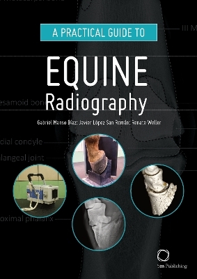
A Practical Guide to Equine Radiography
5m Publishing (Verlag)
978-1-78918-014-5 (ISBN)
This manual is designed to serve as a guide for the clinical veterinarian to perform radiographs on horses. Every area of the horse is included, from the most common (helmet or giblet) to the most difficult (as the spine, thorax or abdomen).
The text is limited to the basic steps a professional should follow to obtain each projection. While the images in each projection consist of a photograph of the patient's positioning, an X-ray example, the same X-ray highlighting the major anatomical structures and a three-dimensional image to demonstrate anatomy.
A Practical Guide to Equine Radiography is designed to accompany the clinical veterinarian either within a hospital setting or out in the field. The book offers an informative step-by-step guide to obtaining high quality radiographs with a focus on image quality, accuracy, consistency and safety.
General principles and equipment are covered before working through the anatomy of the horse with separate chapters devoted to each body region, providing a thorough and detailed picture of the skeletal structure of the horse, making the book an ideal reference for professionals involved with horse health and disease.
Features provided in the book will guide the veterinarian through the stages of taking and interpreting normal radiographs and include:
- Clinical indications of radiographic areas of interest in the horse
- Equipment required
- Preparation and setup guides, supported by photographs
- Projections focusing on radiographic areas of interest, aided by photographs
- x-rays presented with detailed labels, providing a close-up view of skeletal structures
- Three dimensional images demonstrating normal anatomy
A Practical Guide to Equine Radiography is an essential tool for equine practitioners, veterinary students and para-professionals.
Gabriel Manso Díaz, DVM MSc PhD MRCVS, works at the Complutense Veterinary Teaching Hospital in Madrid (Spain) and is a Large Animal Diagnostic Imaging resident at the Royal Veterinary College in London (UK). He splits his time between clinical work in veterinary diagnostic imaging and clinical research, with a particular emphasis on equine head, spinal and abdominal imaging.
Javier López San Román educated at Universidad Complutense de Madrid. Obtained his Ph.D. in equine surgery in 1996. He is heavily involved in teaching veterinary students (general surgery, equine medicine and surgery, and lameness diagnosis), graduate students (sedation and analgesia in the equine patient) and is Associate Professor at the Department of Animal Medicine and Surgery at the Universidad Complutense (Madrid) where he teaches general and special equine surgery. He is Diplomate of the European College of Veterinary Surgeons and actually the Head of the Large Animal Hospital at the Complutense Veternary Teaching Hospital. His research interests include surgical diseases, sedation and analgesia and gait analysis.
After graduating from the University of Munich, Renate Weller spent a year in the US before she returned to Germany to work in equine practice. She then became a senior clinical research scholar in large animal diagnostic imaging at the Royal Veterinary College (RVC). After this she joined the Institute of Veterinary Anatomy in Munich, where she completed her Dr.Vet.Med. thesis on comparison of different imaging modalities in the diagnosis of head disorders in the horse. Following this she spent two years in California before returning to the RVC to do a PhD in the Structure and Motion Laboratory investigating the effect of conformation on locomotor biomechanics in the horse. Since 2005 Renate has been employed at the RVC dividing her time between clincal work in large animal diagnostic imaging and research in imaging, locomotor biomechanics and veterinary education.
General principles
X-ray equipment and radiation safety in equine practice
Quality assessment
Foot
Pastern
Fetlock
Metacarpus/Metatarsus
Carpus
Elbow
Shoulder
Tarsus
Stifle
Pelvis
Head
Cervical
Back
Thorax
Abdomen
| Erscheinungsdatum | 01.03.2019 |
|---|---|
| Zusatzinfo | 325 colour pictures |
| Verlagsort | Sheffield |
| Sprache | englisch |
| Maße | 210 x 297 mm |
| Gewicht | 867 g |
| Einbandart | gebunden |
| Themenwelt | Veterinärmedizin ► Klinische Fächer ► Bildgebende Verfahren |
| Veterinärmedizin ► Pferd ► Bildgebende Verfahren | |
| Schlagworte | Bildgebende Verfahren (Veterinärmedizin) • Pferde; Veterinärmedizin • Radiologie (Veterinärmedizin) |
| ISBN-10 | 1-78918-014-7 / 1789180147 |
| ISBN-13 | 978-1-78918-014-5 / 9781789180145 |
| Zustand | Neuware |
| Haben Sie eine Frage zum Produkt? |
aus dem Bereich


